Delhi | Kolkata
Bangalore | Chennai
Heart Surgery in India
Importance of Heart
Our heart beats 100,000 times a day, pushing 5,000 gallons of blood through our body every 24 hours. It supplies oxygen and nutrient rich blood to our tissues and transports waste away.
The human heart comprises of a pair of atria which accepts deoxygenated blood and drives it into a pair of ventricles which pump blood into the vessels. The right atrium receives the systemic blood from the superior and inferior vena cava which are relatively short in oxygen and pumps it into the right ventricle which propels it into the pulmonary circuit, lungs. Interchange of oxygen and carbon dioxide takes place in the lungs, and blood high in oxygen returns to the left atrium which pumps blood into the left ventricle which in turn pumps blood into the aorta and the remnants of the systemic circuit. The septa are the dividers that separate the chambers of the heart.
Heart surgery is done to rectify problems with the heart. Heart-related problems do not always involve surgery. Sometimes they can be addressed with lifestyle modifications, medicines or nonsurgical procedures. For example, catheter ablation uses energy to make small scars in your heart tissue to prevent abnormal electrical signals from going through your heart. Coronary angioplasty is a minimally invasive procedure in which a stent is introduced into a narrowed or blocked coronary artery to hold it open. However, surgery is often needed to solve problems such as heart failure, plaque build-up that partly or fully blocks blood flow in a coronary artery, defective heart valves, dilated or diseased major blood vessels (such as the aorta) and irregular heart rhythms.
Types of Heart Surgery
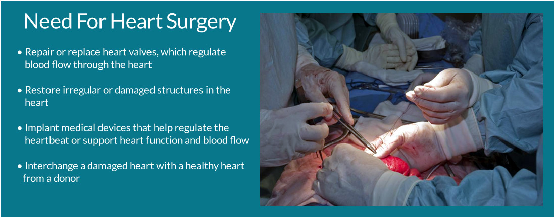
The results of heart surgery in adults often are excellent. Heart surgery can reduce symptoms, improve quality of life, and improve the chances of survival.
Coronary artery bypass grafting (CABG)
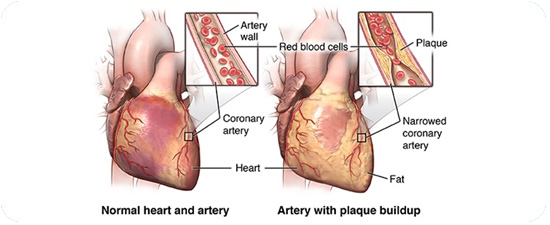
In CABG — the most common type of heart surgery — the surgeon takes a fit artery or vein from somewhere else in your body and links it to supply blood past the blocked coronary artery. The grafted artery or vein go around the blocked part of the coronary artery, forming a fresh path for blood to flow to the heart muscle. Often, this is done for more than one coronary artery in the course of the same surgery. CABG is occasionally referred to as heart bypass or coronary artery bypass surgery.
Heart valve repair or replacement.
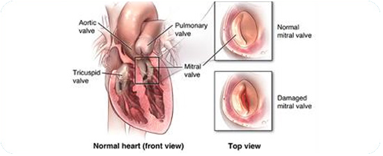
Surgeons either repair the valve or substitute it with an artificial valve or with a biological valve made from pig, cow or human heart tissue. One repair alternative is to insert a catheter through a large blood vessel, lead it to the heart and inflate and deflate a small balloon at the tip of the catheter to expand a narrow valve.
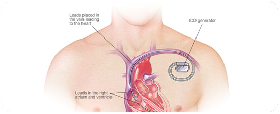
Insertion of a pacemaker or an implantable cardioverter defibrillator (ICD).
Medicine is usually the first treatment option for arrhythmia, a situation in which the heart beats too fast, too slow or with an irregular rhythm. If the medicine does not work, a surgeon may graft a pacemaker under the skin of the chest or abdomen, with wires that connect it to the heart chambers. The device uses electrical pulses to regulate the heart rhythm when a sensor perceives that it is abnormal. An ICD works the same, but it directs an electric shock to restore a normal rhythm when it perceives a dangerous arrhythmia.
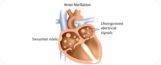
Maze surgery
The surgeon makes a pattern of scar tissue within the upper chambers of the heart to transmit electrical signals along a controlled path to the lower heart chambers. The surgery blocks the stray electrical signals that cause atrial fibrillation — the most common type of serious arrhythmia.

Aneurysm repair
A weak division of the artery or heart wall is replaced with a patch or graft to repair a balloon-like bulge in the artery or wall of the heart muscle.
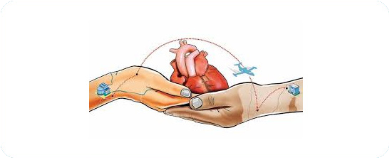
Heart transplant
The diseased heart is detached and substituted with a healthy heart from a deceased donor.
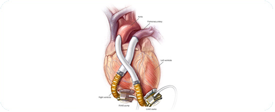
Insertion of a ventricular assist device (VAD) or total artificial heart (TAH).
A VAD is a mechanical pump that maintains heart function and blood flow. A TAH substitutes the two lower chambers of the heart.
In addition to these surgeries, a minimally invasive substitute to open-heart surgery that is becoming more common is trans-catheter structural heart surgery. This encompasses guiding a long, thin, flexible tube called a catheter to your heart through blood vessels that can be gained access to from the groin, thigh, abdomen, chest, and neck or collar bone. A small incision is required. This type of surgery includes trans-catheter aortic valve implantation to substitute a faulty aortic valve with a valve made from animal tissue.
Specialists Involved
Your primary care doctor, a cardiologist, and a cardiothoracic (KAR-de-o-tho-RAS-ik) surgeon will consult with you to choose whether you need heart surgery.
A cardiologist focuses in diagnosing and treating heart problems. A cardiothoracic surgeon concentrates on surgery on the heart and lungs.
These doctors will discuss with you and do tests to study about your overall health and your heart problem.
Medical Evaluation
Your doctors will talk with you about:
- The kind of heart problem you have and it’s indications and for how long you have had those symptoms.
- Your previous treatment of heart problems, including surgeries, dealings, and medicines.
- Genetic history of heart problems.
- Indications of other health problems, such as diabetes or high blood pressure.
- Your age and overall health.
- You also may have blood tests, such as a complete blood count, a lipoprotein panel (cholesterol test), and other tests as required.
Diagnostic Tests
Tests are done to have greater idea about the heart problem and your overall health. This helps your doctor decide whether there is a need for heart surgery, the type of surgery you need, and when it needs to be done.
EKG (Electrocardiogram)
An EKG is a trouble-free, non-invasive test that records the heart's electrical activity. "Non-invasive" means that no surgery is done and no instruments are injected into your body. The test indicates how fast the heart is beating and its rhythm (stable or irregular). An EKG also registers the strength and timing of electrical signals as they pass through the heart. An EKG can show signs of heart impairment due to CHD and signs of a prior or recent heart attack.
Stress Test
Some heart difficulties are easier to make a diagnosis when your heart is working hard and beating fast. During stress testing, you keep fit to make your heart work hard and beat fast. If you can't keep fit, you may be given medicine to increase your heart rate. As part of the test, your blood pressure is tested and an EKG is done.
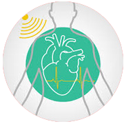
Echocardiography
Echocardiography (echo) is a trouble-free, non-invasive test. This test uses sound waves to generate a moving picture of your heart. Echocardiography shows the dimensions and shape of your heart and how well your heart chambers and valves are functioning. The test also can show areas of poor blood flow to your heart, areas of heart muscle that aren't working well, and earlier injury to your heart muscle caused by poor blood flow.
Coronary Angiography
Coronary Angiography is a test that uses dye and special X-rays to show the interiors of your coronary arteries. To get the dye into your coronary arteries, your doctor will use a process called cardiac catheterization. A thin, flexible tube called a catheter is placed into a blood vessel in your arm, groin (upper thigh), or neck. The tube is eased into your coronary arteries, and the dye is released into your blood. Special X-rays are in use while the dye is flowing through the coronary arteries. These X-rays are called angiograms. The dye helps doctor study blood flow through the heart and blood vessels. This helps your doctor find blockages that can cause a heart attack.

Aortogram
An aortogram is an angiogram of the aorta. The aorta is the chief artery that conveys blood from your heart to your body. An aortogram may show the place and size of an aortic aneurysm.
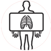
Chest X-Ray
A chest x ray produces representations of the constructions inside your chest, such as your heart, lungs, and blood vessels. This test gives your doctor facts about the size and shape of your heart. A chest x ray also shows the location and shape of the large arteries around your heart.
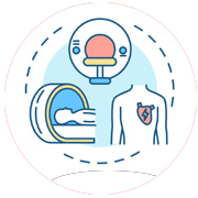
Cardiac Computed Tomography Scan
A cardiac computed tomography (to-MOG-rah-fee) scan, or cardiac CT scan, is a simple test that uses an X-ray machine to take clear, thorough pictures of the heart. Occasionally an iodine-based dye (contrast dye) is inserted into one of your veins during the examination. The contrast dye highlights your coronary (heart) arteries on the X-ray pictures. This type of CT scan is called a coronary CT angiography, or CTA. A cardiac CT scan can display whether plaque is contracting your coronary arteries or whether you have an aneurysm. A CT scan also can find difficulties with the heart's function and valves.
Cardiac Magnetic Resonance Imaging
Magnetic resonance imaging (MRI) is a safe, non-invasive test that uses magnets, radio waves, and a computer to generate pictures of your organs and tissues. Cardiac MRI generates images of your heart as it is beating. The computer makes both still and moving pictures of your heart and major blood vessels. Cardiac MRI shows the structure and function of your heart. This test can show the size and location of an aneurysm.
Risks
Heart surgery has many hazards, even though its results often are exceptional. Risks include:
- - Bleeding.
- - Infection, fever, swelling, and other indications of inflammation.
- - A response to the medicine used to make you sleep during the surgery.
- - Arrhythmias (irregular heartbeats).
- - Injury to tissues in the heart, kidneys, liver, and lungs.
- - Stroke, which may cause temporary or enduring damage.
- - Death.
- - Memory loss and other problems, such as problems directed or thinking clearly, may occur in some people.
These problems are more probable to affect older patients and women. These issues often mend within 6–12 months of surgery.
In overall, the risk of problems is greater if heart surgery is done in an emergency situation (for example, during a heart attack). The danger is also higher if you have other diseases or situations, such as diabetes, kidney disease, lung disease, or peripheral artery disease (P.A.D.)
Before Heart Surgery
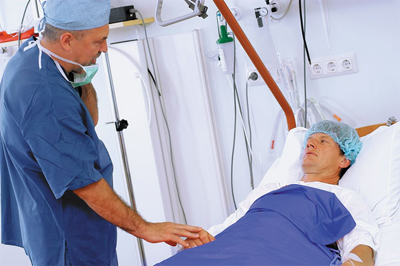
There are numerous types of heart surgery. Some people wisely plan their surgeries with their doctors. They know exactly when and how their surgeries will be planned. Other people need immediate heart surgery. If you're having a planned surgery, your doctors will meet and explain the procedure. You might be admitted to the hospital the afternoon or morning before your surgery.
You may have some tests before the surgery, such as an EKG (electrocardiogram), chest X-ray, or blood tests. An intravenous (IV) line will be positioned into a blood vessel in your arm or chest to give you liquids and medicines.
Before the surgery, the patient is moved to the operating room. The patient is given medicine so that they might fall asleep and not feel any pain during the surgery.
During Heart Surgery
Traditional Open-Heart Surgery - For this category of surgery, the patient is given medicine to help you fall asleep. A doctor will record the heartbeat, blood pressure, oxygen levels, and breathing during the surgery. A breathing tube is placed in the lungs through the throat. The tube connects to a ventilator. The surgeon makes a 6- to 8-inch incision (cut) down the centre of the chest wall. Then, the doctor cuts your breastbone and open your rib cage to reach your heart. During the surgery, the patient receives medicine to thin their blood and keep it from clotting. A heart-lung bypass machine is attached to the heart. The machine controls the heart's pumping action and moves blood away from the heart. A specialist monitors the heart-lung bypass machine. The machine allows the surgeon to operate on a heart that isn't beating and that doesn't have blood flowing through it.
Heart-Lung Bypass Machine – The patient is given medicine to stop the heartbeat once the patient is connected to the heart-lung bypass machine. A tube is placed in the heart to transfer blood to the machine. The machine the removes carbon dioxide from the blood, adds oxygen, and then pumps the blood back into the body. The surgeon the insert tubes into the chest to drain fluid. Once machine starts to work, the surgeon repairs the heart problem. After the surgery the surgeon restores blood flow to the heart. Usually, the heart starts beating again on its own. Occasionally mild electric shocks are used to revive the heart. Once the heart has started beating again, the surgeon removes the tubes and stops the machine. Medicine is then given to allow the blood to clot again. The surgeon then uses wires to close the breastbone. The wires stay in the body permanently. After the breastbone heals, it recovers to be as strong as it was before the surgery. Stitches or staples are used to close the skin incision. The breathing tube is then removed when the patient is able to breathe without it.
Off-Pump Heart Surgery - Off-pump heart surgery is like traditional open-heart surgery because the chest bone is exposed to reach the heart. However, the heart isn't halted, and a heart-lung bypass machine isn't utilised. Instead, the surgeon steadies the heart with a mechanical device so he or she can work on it. The heart will continue to pump blood to your body./
Minimally Invasive Heart Surgery - For this type of heart surgery, the surgeon makes small incisions in the side of the chest between the ribs. These cuts can be as small as 2–3 inches. The surgeon inserts surgical tools through these cuts. A tool with a small video camera at the tip is also inserted through an incision. The tool allows the surgeon to see inside the body. Some kinds of minimally invasive heart surgery use a heart-lung bypass machine and others don't.
Recovery in the Hospital
After the surgery the patient spends a day or more in the hospital's intensive care unit (ICU), dependent on the category of heart surgery. An intravenous (IV) needle is sometimes implanted in the blood vessel in the arm or chest to give fluids until the patient is ready to drink on their own. The health care team might give an extra oxygen through a face mask or nasal prongs that fit just inside the nose. They will remove the mask or prongs when the patient no longer needs them. After leaving the ICU, the patient is moved to another part of the hospital for several days before they can go home.
Recovery at Home
Different people respond differently to heart surgery. The recovery at home depends on what kind of heart problem and surgery the patient had. The doctor will guide on how to:
• Care for the mending incision(s)
• Identify signs of infection or other impediments
• Deal with the after-effects of surgery
The patient also receives information about follow-up appointments, medicines, and situations when there is a need to call the doctor right away.
After-effects of heart surgery are common. They may comprise of muscle pain, chest pain, or swelling, etc. Other after-effects may include loss of appetite, sleeping problems, constipation, and mood swings and depression. After-effects usually go away after some time.
Ongoing Care
Ongoing care after the surgery includes check-ups with your doctor. During these visits, blood tests, an EKG (electrocardiogram), echocardiography, or a stress test might be conducted. Such tests will show how the heart is functioning after the surgery. After some types of heart surgery, there is a need to take a blood-thinning medicine. The doctor will perform routine tests to make sure that the patient is getting the right amount of medicine. The doctor also may advise routine changes and medicines to help stay healthy. Lifestyle changes may include quitting smoking, diet change, physical activity, and decreasing and dealing with stress. The doctor may also refer the patient to cardiac rehabilitation. Cardiac rehab consists of exercise training, education on heart healthy living, and therapy to reduce stress and help in recovery.
* Conditions Apply

